Microstructural features of water fern Salvinia natans (L.) All. organ surfaces
Abstract
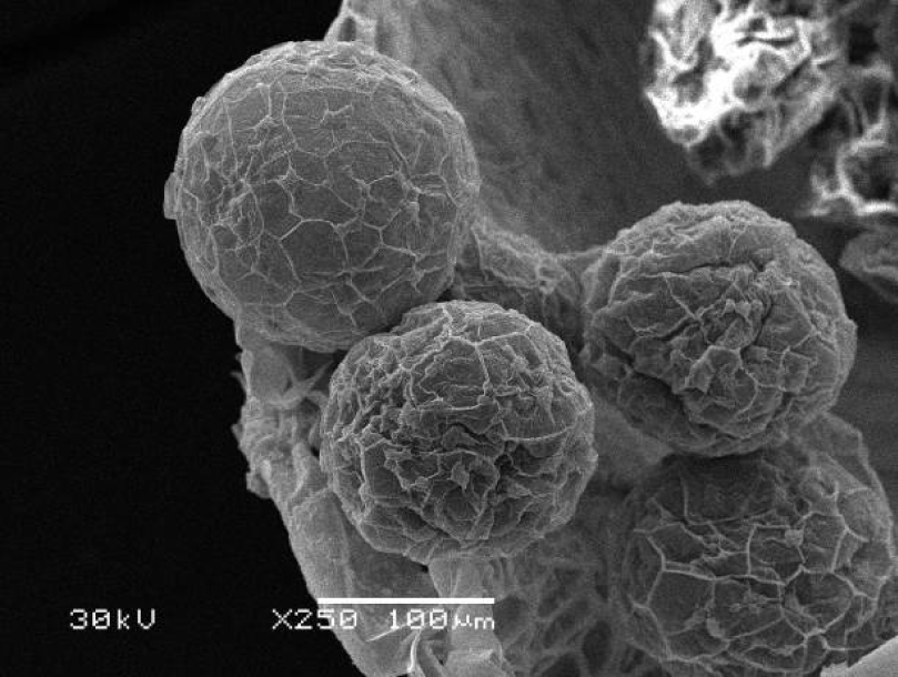
The microstructure of the organs surface of the water fern Salvinia natans (L.) All. has been studied under scanning electron microscope. It was established that the existence on the border between air and water environments is suported by specific microstructure of floating leaves. The adaxial side of floating leaves has well-developed cuticle and stomata placed below the level of epidermis, while abaxial surface of such leaves and submerged modified leaves are characterized by ultra-thin cell walls of the epidermis and numerous filamentous trichomes. We calculated number of stomata per unit area of leaves and the average diameter of stomata. It is claimed that the structure of wall of the sporocarp promotes the passage of the annual summer-green rhythm. Sporocarp provides diving of mega- and microsporangia to the bottom of the water reservoir in autumn and their raising on the water surface in the spring after destruction of its walls.
References
Дубина Д.В., ШелягСосонко Ю.Р., Жмуд О.І., Дворецький Т.В., Дзюба Т.П. 2003. Дунайський біосферний заповідник. Рослинний світ. Фітосоціоцентр, Київ.
Клименко О.М. 2012. Морфолого-анатомічні особливості наземних, плаваючих та придонних листків Nuphar lutea (L.) Smith. Mod. Phytomorphol. 2: 59–62.
Холодный Н.Г. 1956. О метаморфозе пластид в волосках подводных листьев у Salvinia natans. В кн.: Холодный Н.Г. Избранные труды в 3 т. Т. 1. Работы по физиологии растений. Издательство АН УССР, Киев.
Чорна Г.А. 2001. Рослини наших водойм (атлас-довідник). Фітосоціоцентр, Київ.
Meusel H., Jäger E., Weinert E. 1965. Vergleichende Chorologie der zentraleuropäischen Flora. VEB Gustav Fischer Verlag, Jena.
Nagalingum N.S., Schneider H., Pryer K.M. 2006. Comparative morphology of reproductive structures in heterosporous water ferns and a reevaluation of the sporocarp. Int. J. Plant Sci. 167: 805–815.
Stern K.R., Jansky S., Bidlack J.E. 2003. Introductory plant biology. McGraw-Hill, New York.

This work is licensed under a Creative Commons Attribution-NonCommercial-NoDerivatives 4.0 International License.
The journal is licensed by Creative Commons under BY-NC-ND license. You are welcome and free to share (copy and redistribute the material in any medium or format) all the published materials. You may not use the material for commercial purposes. You must give appropriate credit to all published materials.
The journal allow the author(s) to hold the copyrights and to retain publishing rights without any restrictions. This is also indicated at the bottom of each article.