Micromorphology and ultrastructure of the floral nectaries of Viola odorata L. (Violaceae)
Abstract
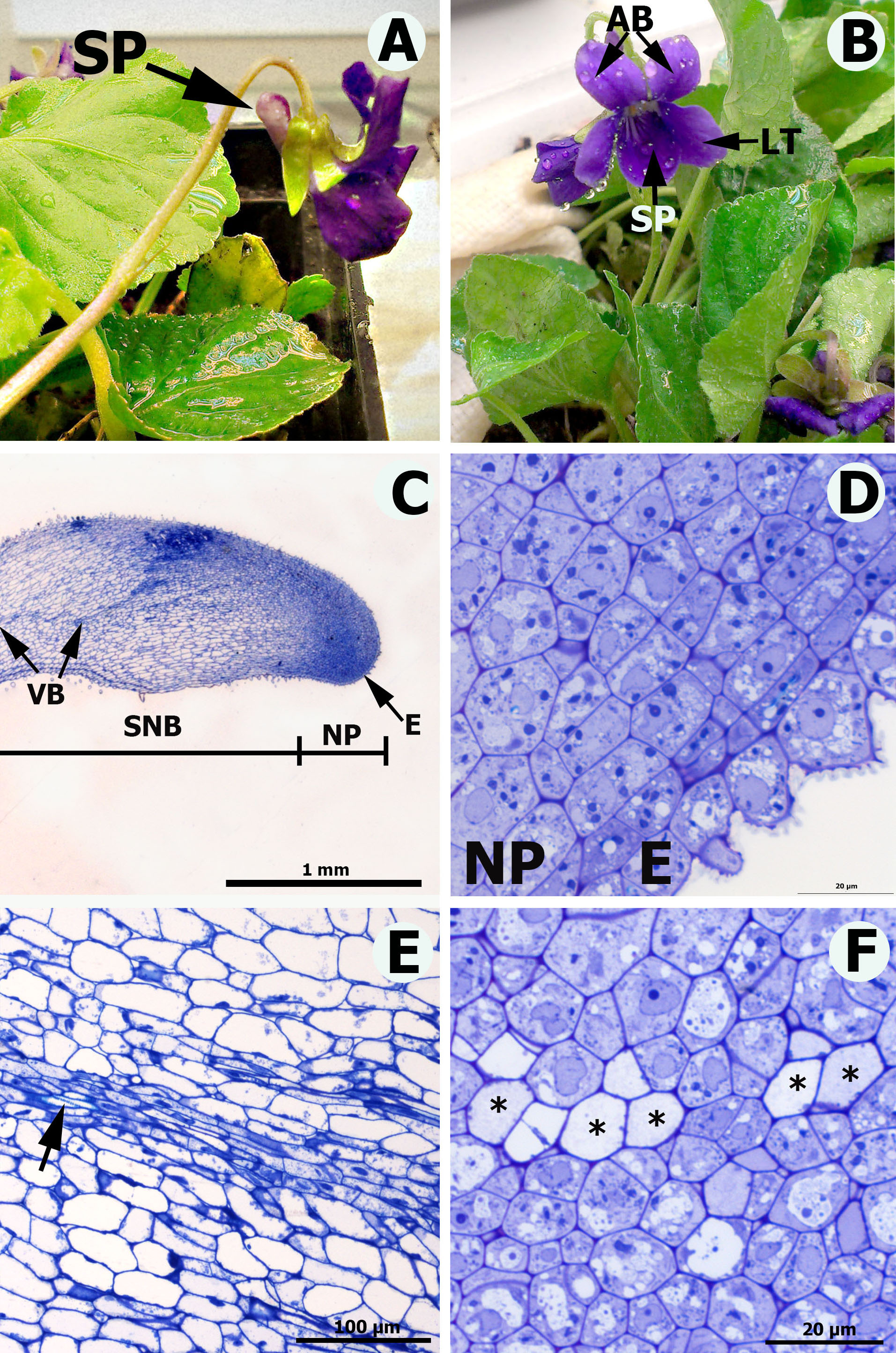
In Viola odorata two inferior anthers have connective appendages (nectaries) projecting into the corolla spur. Nectaries are approx. 4 mm long, elongate, with the top of the nectary bending to the lateral wall of the spur. In the top part and in the abaxial surface of middle part of the nectary all cells have papillae. Nectar is secreted through the modified stomata distributed mainly in the top of nectary The nectary consists of single-layered epidermis, nectary parenchyma and subnectary parenchyma. Features of the nectary parenchyma cells, like dense cytoplasm containing numerous mitochondria and large nuclei, are connected with high metabolic cell activity. The vascularization includes both phloem and xylem. A slight amount of starch in the nectary cells, the profusion of plasmodesmata connecting secretory cells and the presence of vascular bundles suggest that sugars secreted in the nectar were delivered by the phloem sap.
References
Coutinho Í.A.C., Francino D.M.T., Azevedo A.A., Alves Meira R.M.S. 2012. Anatomy of the extrafloral nectaries in species of Chamaecrista section Absus subsection Baseophyllum (Leguminosae, Caesalpinioideae). Flora 207 (6): 427–435. doi: 10.1016/j.flora.2012.03.007
Durkee L.T. 1982. The floral and extrafloral nectaries of Passiflora. II. The extrafloral nectary. Am. J. Bot. 69: 1420–1428.
Fahn A. 1979. Ultrastructure of nectaries in relation to nectar secretion. Am. J. Bot. 66 (8): 977–985.
Fahn A. 1988. Secretory tissues in vascular plants. New Phytol. 108: 229–257.
Fahn A. 2000. Structure and function of secretory cells. Adv. Bot. Res. 35: 37–75.
Konarska A. 2011. Flower nectary structure in Cornus alba L. Plant Syst. Evol. 291: 1–6.
Kowalkowska A.K., Kozieradzka-Kiszkuno M., Turzyński S. 2014. Morphological, histological and ultrastructural features of osmophores and nectary of Bulbophyllum wendlandianum (Kraenzl.) Dammer (B. section Cirrhopetalum Lindl., Bulbophyllinae Schltr., Orchidaceae). Plant Syst. Evol. 301: 609–622.
Nepi M. 2007. Nectary structure and ultrastructure. In: Nicolson S.W., Nepi M., Pacini E. (eds), Nectaries and nectar: 129–166. Springer, Dordrecht.
Pacini E., Nepi M., Vesprini J.L. 2003. Nectar biodiversity: a short review. Plant Syst. Evol. 238: 7–22.
Paiva E.A.S. 2009. Ultrastructure and post-floral secretion of the pericarpial nectaries of Erythrina speciosa (Fabaceae). Ann. Bot. 104: 937–944.
Paiva E.A.S., Machado S.R. 2008. The floral nectary of Hymenaea stigonocarpa (Fabaceae, Caesalpinioideae): structural aspects during floral development. Ann. Bot. 101: 125–133.
Spurr A.R. 1969. A low viscosity epoxy resin embedding medium for electron microscopy. J. Ultrastructure Res. 26: 31–43.
Stpiczyńska M., Nepi M., Zych M. 2012. Secretion and composition of nectar and the structure of perigonal nectaries in Fritillaria meleagris L. (Liliaceae). Plant Syst. Evol. 298: 997–1013.
Vassilyev A.E. 2010. On the mechanisms of nectar secretion: revisited. Ann. Bot. 105: 349–354.

This work is licensed under a Creative Commons Attribution-NonCommercial-NoDerivatives 4.0 International License.
The journal is licensed by Creative Commons under BY-NC-ND license. You are welcome and free to share (copy and redistribute the material in any medium or format) all the published materials. You may not use the material for commercial purposes. You must give appropriate credit to all published materials.
The journal allow the author(s) to hold the copyrights and to retain publishing rights without any restrictions. This is also indicated at the bottom of each article.