Comparative rhizome anatomy of some species of Ceradenia L.E. Bishop and Zygophlebia L.E. Bishop (Polypodiaceae, formerly Grammitidaceae) from Madagascar
Abstract
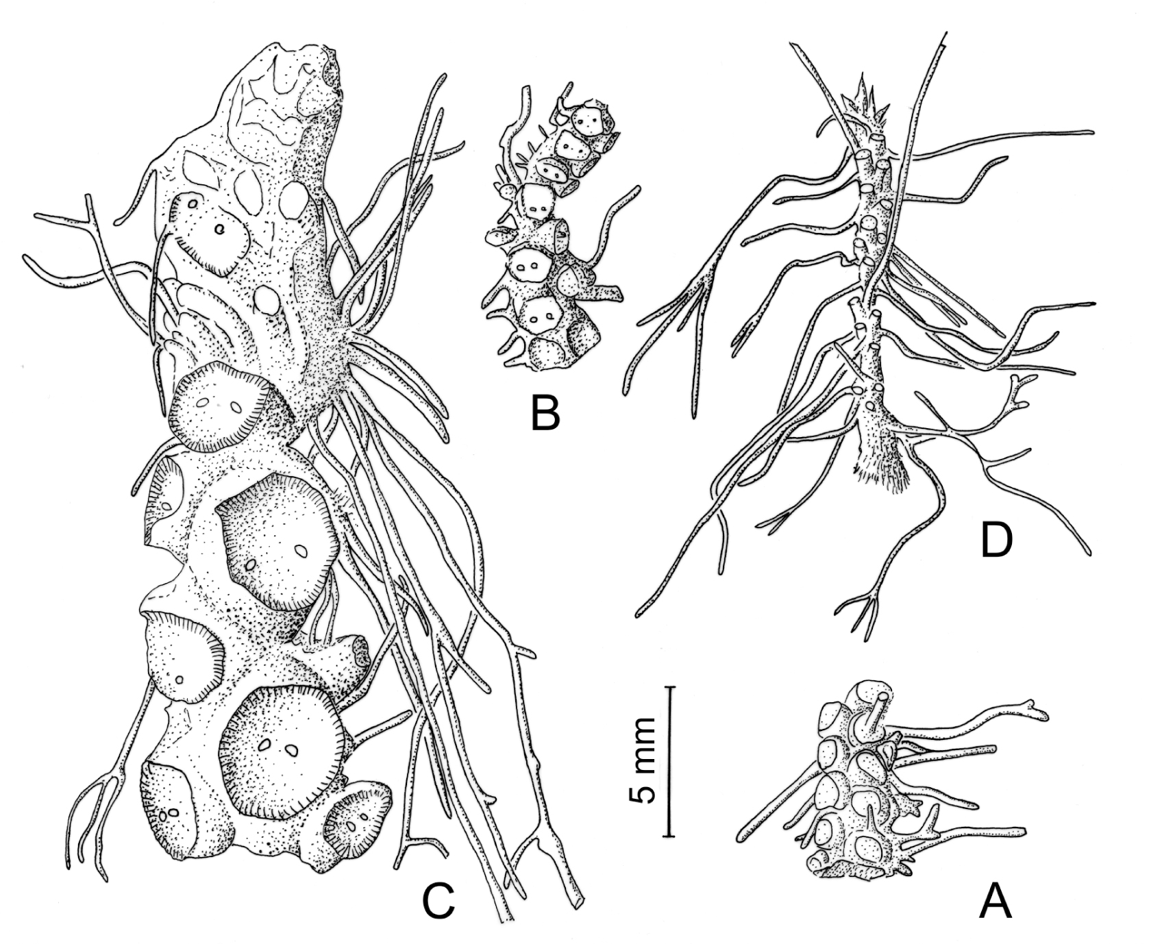
Rhizome anatomy of 2 species of Ceradenia and 2 species of Zygophlebia, mainly Malagasy endemics, was studied in detail, and compared with preliminary results previously achieved in 2 other Zygophlebia species from Madagascar. Stele architecture was reconstructed in each species, with two sketches (a cross section and a splitting-out diagram), thus providing anatomical features and allowing a discussion about their relevance for distinguishing the two genera. Especially, we emphasize that Ceradenia differs from Zygophlebia by lacking any accessory ventral gap, but that character has a value only in combination with the occurrence of whitish waxy hairs in the sori. On the other hand, histology provides few correlated results, due to a narrow sampling, but tracheids lumen appears in transverse section sinuate in Ceradenia, rounded in Zygophlebia. Rhizotaxis was analyzed, revealing a more or less helical insertion of roots, along meridians in definite number, mainly crowded at the ventral rhizome side, and whose divergence angle seems to be little altered by environmental constraints. Phyllopodial divergence angles are constant too. All these geometrical data may be useful for characterizing species, or species groups, but not at a generic level. They appear however not tightly correlated to rhizome size and might have an adaptive significance.
References
Bishop L.E. 1989. Zygophlebia, a new genus of Grammitidaceae. Am. Fern J. 79: 103–118.
Deroin T., Rakotondrainibe F. 2010. Comparative rhizome anatomy of some species of Ceradenia and Zygophlebia (Polypodiaceae, formerly Grammitidaceae) from Madagascar. In: Jeannoda V.H., Razafimandimbison S.G., De Block P. (eds), XIXth AETFAT Congress Madagascar. Scripta Bot. Belg. 46: 130.
Gerlach D. 1984. Botanische Mikrotechnik. Ed. 3, Thieme, Stuttgart.
Hovenkamp P. 1990. The significance of rhizome morphology in the systematics of the polypodiaceous ferns (sensu stricto). Am. Fern J. 80: 33–43.
Lachmann J.-P. 1889. Contribution à l’Histoire Naturelle de la Racine des Fougères. Thesis, Univ. Paris, Ser. A, 116, 653. Association typographique, Lyon.
Leroux O., Bagniewska-Zadworma A., Rambe S.K., Knox J.P., Marcus S.E., Bellefroid E., Stubbe D., Chabbert B., Habrant A., Claeys M., Viane R.L.L. 2011. Non-lignified helical cell thickening in root cortical cells of Aspleniaceae (Polypodiales): histology and taxonomical significance. Ann. Bot. 107: 195-207.
Ogura Y. 1938. Anatomie der Vegetationsorgane der Pteridophyten in K. Linsbauer, Handbuch der Pflanzenanatomie. II. Abteilung, Band VII, Teil 2: Archegoniaten B, Borntraeger, Berlin.
Rakotondrainibe F., Deroin T. 2006. Comparative morphology and rhizome anatomy of two new species of Zygophlebia (Grammitidaceae) from Madagascar and notes on the generic circumscription of Zygophlebia and Ceradenia. Taxon 55: 145–152.
Srivastava A., Chandra S. 2009. Structure and organization of the rhizome vascular system of four polypodium species. Am. Fern J. 99: 182–193.
Sundue M.A. 2010. A morphological cladistic analysis of Terpsichore (Polypodiaceae). Syst. Bot. 35: 716–729.
Sundue M.A., Parris B.S., Ranker T.A., Smith A.R., Fujimoto E.L., Zamora-Crosby D., Morden C.W., Chiou W.-L., Chen C.-W., Rouhan G., Hirai R.Y., Prado J. 2014. Global phylogeny and biogeography of grammitid ferns (Polypodiaceae). Mol. Phylogenet. Evol. 81: 195–206.
Tansley M.A. 1907. Lectures on the evolution of the filicinean vascular system. VI. The Evolution of dictyostely. Polycycly. New Phytologist 6: 187–203.
Wetter C. 1952. Untersuchungen über Anordnung und Anlegung der Wurzeln bei leptosporangiaten Farnen. Abhandlungen der mathematische-naturwissenschaftliche Klasse, Akademie für Wissenschaft und Literatur, Mainz. Jahrgang 1952 (1): 26–84.


This work is licensed under a Creative Commons Attribution-NonCommercial-NoDerivatives 4.0 International License.
The journal is licensed by Creative Commons under BY-NC-ND license. You are welcome and free to share (copy and redistribute the material in any medium or format) all the published materials. You may not use the material for commercial purposes. You must give appropriate credit to all published materials.
The journal allow the author(s) to hold the copyrights and to retain publishing rights without any restrictions. This is also indicated at the bottom of each article.


