Structural organization of Cobeae scandens Cav. nectaries
Abstract
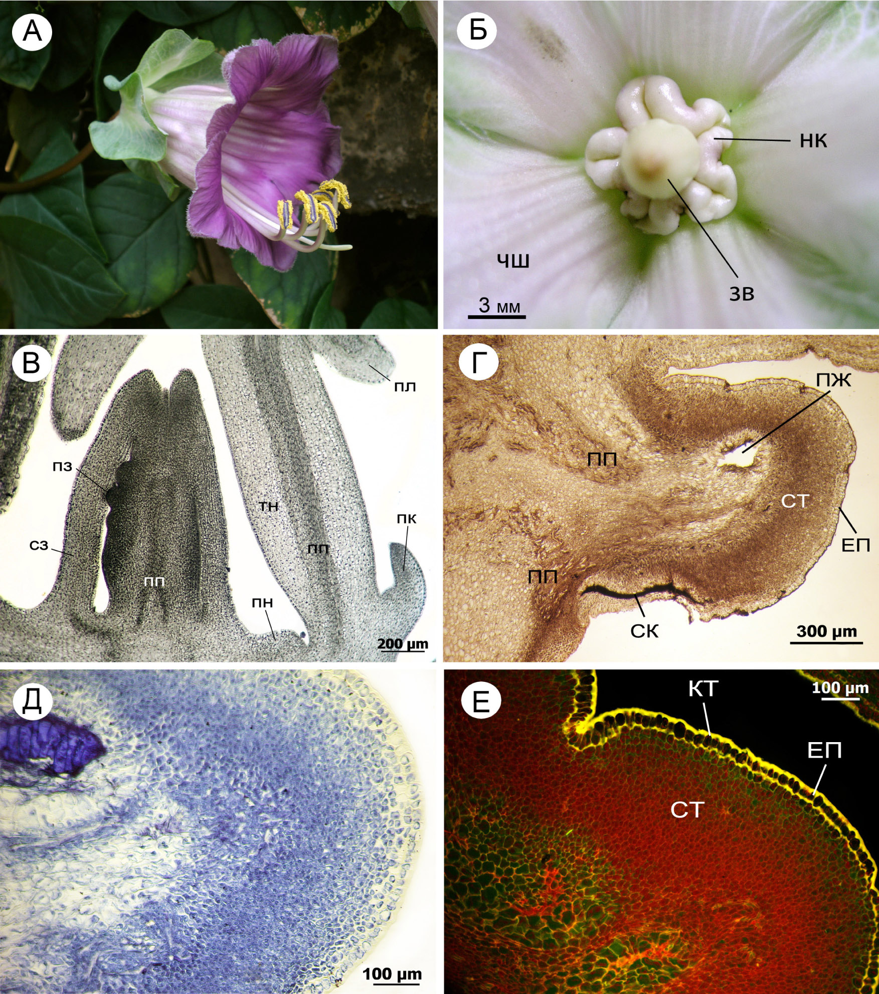
It has ascertained, that Cobea scandens Cav., which introducted in the National botanic garden of M.M. Grishka NAN of Ukraine, has secretory tissues, which develop and function normally. In anatomical structure of C. scandens nectaries five structure-functional areas were founded. In the cells of secretory area a lot of RNA and proteins were detected. Intrastaminal nectaries have many common morpho-anatomical features with stamens. It is support of M. Dawson opinion (see Тахтаджян 1966) about staminal origin of C. scandens nectaries.
References
Тахтаджян А.Л. 1966. Система и филогения цветковых растений: 422–424 Наука, Москва – Ленинград.
Тахтаджян А.Л. (ред.). 1981. Жизнь растений. Т. 5 (2): 390–392. Просвещение, Москва.
Фурст Г.Г. 1979. Методы анатомо-гистохимического исследования растительных тканей: 40–65.Наука, Москва.

This work is licensed under a Creative Commons Attribution-NonCommercial-NoDerivatives 4.0 International License.
The journal is licensed by Creative Commons under BY-NC-ND license. You are welcome and free to share (copy and redistribute the material in any medium or format) all the published materials. You may not use the material for commercial purposes. You must give appropriate credit to all published materials.
The journal allow the author(s) to hold the copyrights and to retain publishing rights without any restrictions. This is also indicated at the bottom of each article.