The model of areolae distribution and surface area of pores located on the valves of centric diatoms Coscinodiscus Ehr. and Thalassiosira Cl.
Abstract
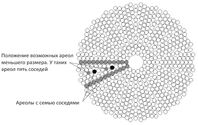
Chemical substances exchange between microalgae and environment occurs through the pores on the surface of its coat. Therefore, characteristics of substance flow are related to the total area of the pores of microalgae. Pores of centric diatoms are arranged in groups on the siliceous plates that cover the areolae of valves. The mathematical model that simulates the radial areolae arrangement on the valves of centric diatoms and on the basis of estimated number of areolae calculates the total area of the diatom pores is proposed. On the example of two diatom species Coscinodiscus sp. and Thalassiosira eccentrica it was shown that their pores occupy 4-8% and 7-14% of total valve surface respectively. These values do not depend on the valve diameter and number of its areolae but are equivalent to the porosity of areolae of these species. It is supposed that about 10% of valve surface takes part in substance exchange and 100% of valve surface absorbs sunlight. Therefore substance flow through the valve is related to the energy flow (sunlight) as 1:10. It is suggested to use the ratio between total area of microalgae surface and area of its pores for the characteristics of microalgae productivity.
References
Bentley K., Cox E.J., Bentley P.J. 2005. Nature’s batik: a computer evolution model of diatom valve morphogenesis. J. Nanosc. Nanotech. 5: 1–10.
Bukhtiyarova L.N. 2009. Frustule functions and functional morphology of Bacillariophyta. Альгология 19 (3): 321–331.
De Stefano L., Rea I., Rendina I., De Stefano M., Moretti L. 2007. Lensless light focusing with centric marine diatom Coscinodiscus walesii. Opt. Express 15: 18082–18088.
Fryxell G.A., Ashworth T.K. 1988. The diatom genus Coscinodiscus Ehrenberg: characters having taxonomic value. Bot. Marina 31: 359–374.
Fuhrmann T., Landwehr S., El Rharbi-Kucki M., Sumper M. 2004. Diatoms as living photonic crystals. Applied Physics B 78: 257–260.
Hicks Y.A., Marshall D., Rosin P.L., Martin R.R., Mann D.G., Droop S.J.M. 2006. A model of diatom shape and texture for analysis, synthesis and identification. Mach. Vision Appl. 17: 297–307.
Lenoci L., Camp P.J. 2008. Diatoms structures templated by phase-separated fluids. Langmuir 24: 217–223.
Longuet-Higgins M.S. 2001. Geometrical constraints on the development of a diatom. J. Theoretical Biology 210: 101–105.
Losic D., Pillar R.J., Dilger T., Mitchel J.G., Voelcker N.H. 2007. Atomic force microscopy (AFM) characterisation of the porous silica nanostructure of two centric diatoms. J. Porous Mater. 14: 61–69.
Noes J., Sumper M., Vukusic P. 2008. Light manipulation in a marine diatom. J. Mater. Res. 23: 3229–3235.
Parkinson J., Brechet Y., Gordon R. 1999. Centric diatom morphogenesis: a model based on a DLA algorithm investigating the potential role of microtubules. Biochim. Biophys. Acta 1452: 89–102.
Willis L., Page K.M., Broomhead D.S., Cox E.J. 2010. Discrete free-boundary reaction-diffusion model of diatom pore occlusion. Plant Ecol. Evol. 143 (3): 297–306.

This work is licensed under a Creative Commons Attribution-NonCommercial-NoDerivatives 4.0 International License.
The journal is licensed by Creative Commons under BY-NC-ND license. You are welcome and free to share (copy and redistribute the material in any medium or format) all the published materials. You may not use the material for commercial purposes. You must give appropriate credit to all published materials.
The journal allow the author(s) to hold the copyrights and to retain publishing rights without any restrictions. This is also indicated at the bottom of each article.