The ultrastructurе of chloroplasts and photosynthetic pigments in floating and submerged leaves of water fern Salvinia natans (L.) All during ontogeny
Abstract
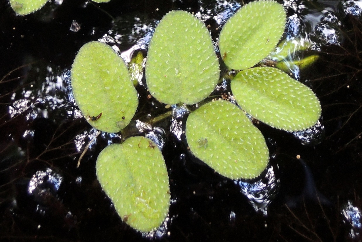
The results of the comparative analysis of chloroplast ultrastructure and analysis of photosynthetic pigments content in floating and submerged leaves of water fern Salvinia natans (L.) All. at the different stages of ontogeny are presented. The ultrastructure of photosynthetic organelles and pigments content are significantly different in floating and submerged leaves. The chloroplasts of parenchymal cells of floating leaves have a well-developed membranous system with many grana and contain many starch grains. Submerged leaves were shown to form smaller chloroplasts with low starch content in the stroma. A smaller number and smaller size of grana complexes in chloroplasts were marked, too. Destructive changes in the photosynthetic membranes of chloroplasts in both types of leaves were observed at the stage of sporocarps formation. The content of photosynthetic pigments in the floating leaves was twice higher than in the submerged leaves, and at the certain stages of ontogeny three times higher. During development of the plant, a content of photosynthetic pigments raised up in the floating leaves. At the stage of sporocarps formation, some reduction of chlorophylls and carotenoids content in submerged leaves occurred. In this article, we discuss the relationship between the identified differences and the functional activity of floating and submerged leaves during growth and development of water fern S. natans.
References
[Andrianova Yu.E., Tarchevskiy I.A. 2000. Chlorophyll and productivity of plants. Nauka, Moscow. (In Russian)]
Дубина Д.В., Шеляг-Сосонко Ю.Р., Жмуд О.І., Дворецький Т.В., Дзюба Т.П. 2003. Дунайський біосферний заповідник. Рослинний світ. Фітосоціоцентр, Київ.
[Dubyna D.В., Shelyag-Sosonko Yu.R., Zhmut O.I., Dvoretsky T.V., Dziuba T.P. 2003. Danube biosphere reserve. Plants. Phytosociocentr, Kyiv. (In Ukrainian)]
Клименко О.М. 2014. Структурно-функціональні аспекти гетерофілії Nuphar lutea (L.) Smith.: Автореф. дис... к-та біол. наук. Київ.
[Klymenko O.M. 2014. Structufal and functional aspects of Nuphar lutea heterophylly (L.) Smith. Thesis of PhD manuscript. Kyiv. (In Ukrainian)]
Кочубей С.М., Бондаренко О.Ю., Шевченко В.В. 2014. Фотосинтез. Т. 1. Структурная организация и функциональные особенности световой фазы фотосинтеза. Логос, Киев.
[Kochubey S.M., Bondarenko O.Yu., Shevchenko V.V. 2014. Photosynthesis. Vol. 1. The structure and functional peculiarities of light phase of photosynthesis. Logos, Kiev. (In Russian)]
Недуха О.М. 2011а. Гетерофілія у рослин. Альтерпрес, Київ.
[Nedukha O.M. 2011а. Heterophylly in plants. Alterpress, Kyiv. (In Ukrainian)]
Недуха О.М. 2011б. Ультраструктурна характеристика клітин та аналіз пігментів плаваючих і підводних листків Trapa natans L. Mod. Phytomorphol. 1: 81–84.
[Nedukha O.M. 2011б. The ultrastructural characteristics of cells and pigments of Trapa natans L. floating and submerged leaves. Mod. Phytomorphol. 1: 81–84. (In Ukrainian)]
Недуха О.М. 2015. Aнатомічна будова та особливості ультраструктури хлоропластів листків деяких гідрофітів. Mod. Phytomorphol. 8: 162–168.
[Nedukha O.M. Anatomy of leaves and chloroplasts ultrastructure of some hydrophytes. Mod. Phytomorphol. 8: 162–168. (In Ukrainian)]
Некрасова Г.Ф, Ронжина Д.А, Коробицына Е.Б. 1998. Формирование фотосинтетического аппарата в период роста погруженного, плавающего и надводного листа гидрофитов. Физиол. раст. 45: 539–548.
[Nekrasova G.F., Ronzhina D.A., Korobitsina E.B. 1998. The photosynthetic system formation during the growth of submerged, floating and overwater leaves of hydrophytes. Plant Physiol. 45: 539–548. (In Russian)]
Некрасова Г.Ф., Ронжина Д.А., Малева М.Г., Пьянков В.И. 2003. Фотосинтетический метаболизм и активность карбоксилирующих ферментов у надводных, плавающих и погруженных листьев гидрофитов. Физиол. раст. 50: 65–75.
[Nekrasova G.F., Ronzhina D.A., Maleva M.G., Pyankov V.I. 2003. The photosynthetic metabolism and activity of the carboxylation enzymes in overwater, floating and submerged leaves of hydrophytes. Plant Physiol. 50: 65–75. (In Russian)]
Холодный Н.Г. 1956. О метаморфозе пластид в волосках подводных листьев у Salvinia natans. В кн: Холодный Н.Г. Избранные труды в 3 т. Т. 1. Работы по физиологии растений. Издательство АН УССР, Киев.
[Kholodny N.G. 1956. About metamorphosis of plastids in hairs of Salvinia natans submerged leaves. In: Kholodny N.G. Selected papers in 3 vol. Vol. 1. The papers about the plant physiology. Publishing of the UkrSSR Academy of Sciences. Kiev (In Russian)]
Чорна Г.А. 2001. Рослини наших водойм (атлас-довідник). Фітосоціоцентр, Київ.
[Chorna G.A. 2001 The plants of our water reservoirs (guidebook). Phytosociocentr, Kyiv. (In Ukrainian)]
Щербатюк М. М., Бриков В. О., Мартин Г. Г. 2015. Підготовка зразків рослинних тканин для електронної мікроскопії (теоретичні та практичні аспекти). Талком, Київ.
[Shcherbatiuk M. M., Brykov V. O., Martyn G. G. 2015. The preparation of plant tissues for electron microscopy (theoretical and practical aspects). Talkom, Kyiv. (In Ukrainian)]
Austin J. R., Frost E., Vidi P.-A., Kessler F., Staehelin L. A. 2006. Plastoglobules are lipoprotein subcompartments of the chloroplast that are permanently coupled to thylakoid membranes and contain biosynthetic enzymes. Plant Cell 18: 1693–1703.
Babenko L., Kosakivska I., Akimov Yu., Klymchuk D., Skaternya T. 2014. Effect of temperature stresses on pigment spectrum, lipoxygenase activity and cell ultrastructure of winter wheat seedling. Genet. Plant Physiol. 4: 117–125.
Barthlott W., Wiersch S., Čolić Z., Koch K. 2009. Classification of trichome types within species of the water fern Salvinia, and ontogeny of the egg-beater trichomes. Botany 87: 830–836.
Bercu R. 2004. Histoanatomy of the leaf of Trapa natans (Trapaceae). Phytol. Balcan. 10: 51–55.
Bercu R. 2006. Anatomical features of the vegetative organs of Salvinia natans (L.) All. (Salviniaceae). USAMVBT Symposium. 5th section: Biology researchers with implications in agriculture: 321–324.
Bréhélin C., Kessler F., Van Wijk K. J. 2007. Plastoglobules: versatile lipoprotein particles in plastids. Trends Plant Sci. 12: 260–266.
Croxdale J.G. 1978. Salvinia leaves. I. Origin and early differentiation of floating and submerged leaves. Can. J. Bot. 56: 1982–1991.
Croxdale J.G. 1981. Salvinia leaves. III. Morphogenesis of the submerged leaf. Can. J. Bot. 59: 2065–2072.
Evert R.F. 2007. Esau’s plant anatomy. 3rd edition. Wiley Interscience, Hoboken, New Jersey.
Lemon G.D., Posluszny U. 1997. Shoot morphology and organogenesis of the aquatic floating fern Salvinia molesta D.S. Mitchell, examined with the aid of laser scanning confocal microscopy. Int. J. Plant Sci. 158: 693–703.
Nielsen S.L. 1993. A comparison of aerial and submerged photosynthesis in some Danish amphibious plants. Aquat. Bot. 45: 27–40.
Рringsheim N. 1863. Zur Morphologie der Salvinia natans. Jahrbuch für wissenschaftliche Botanik. 3: 484–541.
Ryen F.J. 1985. Isolation and characterization of photosynthetically active cells from submerged and floating leaves of the aquatic macrophyte Potamogeton nodosus Poir. Plant Cell Physiol. 26: 309–315.
Sand-Jensen K., Frost-Christensen H. 1999. Plant growth and photosynthesis in the transition zone between land and stream. Aquat. Bot. 63: 23–35.
Spicher L., Kessler F. 2015. Unexpected roles of plastoglobules (plastid lipid droplets) in vitamin K1 and E metabolism. Curr. Opin. Plant Biol. 25:123–129.
Wellburn A. 1994. The spectral determination of chlorophyll a and chlorophyll b, as well as total carotenoids, using various solvents with spectrophotometers of different resolution. J. Plant Physiol. 144: 307–313.
Yang J.P., Dengler N.G., Horton R.F.1987. Heterophylly in Ranunculus flabellaris: The effect of abscisic acid on leaf anatomy. Ann. Bot. 60: 117–125.

This work is licensed under a Creative Commons Attribution-NonCommercial-NoDerivatives 4.0 International License.
The journal is licensed by Creative Commons under BY-NC-ND license. You are welcome and free to share (copy and redistribute the material in any medium or format) all the published materials. You may not use the material for commercial purposes. You must give appropriate credit to all published materials.
The journal allow the author(s) to hold the copyrights and to retain publishing rights without any restrictions. This is also indicated at the bottom of each article.